
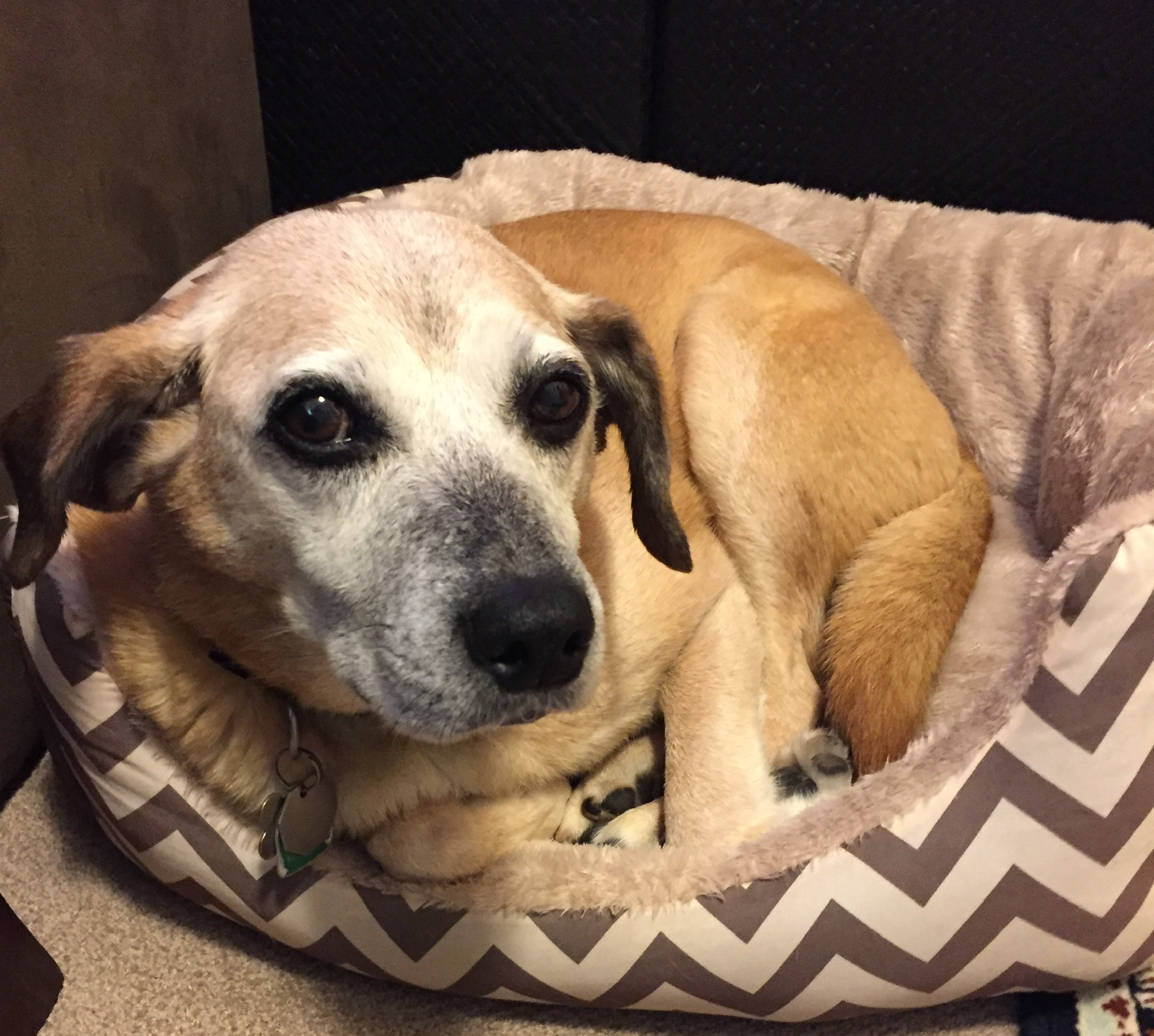
Lily is a 15-year-old athletic hound mix. She has always been very fit and loves going on long hikes with her owners. As Lily has aged, she has shown very little sign of slowing down.
During one of her regular check-ups, her owners told the veterinarian that Lily has slowed down this last year. During Lily’s exam, a routine dental prophylaxis with full mouth x-rays was recommended. Lily also had some orthopedic pain consist with age related osteoarthritis.
It was decided to go ahead with her dental procedure, but prior to this we wanted to make sure Lily was entirely healthy. She had blood tests to check her organ function, electrolytes, blood sugar, red blood cells and platelets. She also had an ECG to check her heart. It was determined that Lily was safe to undergo anesthesia.
For dogs and cats all dental procedures must be done under anesthesia. Animals need to be anesthetized so the veterinarian can perform a thorough oral exam. Each tooth is given a detailed exam and full mouth x-rays are done to ensure the entire tooth is evaluated. In dogs and cats the roots make up over 50% of the tooth, so without x-rays we are not evaluating over half of each tooth.
Lily was anesthetized and closely monitored during her procedure. A detailed oral exam was performed and each tooth examined. Once this was done, Lily had a deep periodontal cleaning and full mouth x-rays. Her x-rays showed some surprising findings, there were multiple teeth that were resorbing. Without doing x-rays we would have missed this painful condition. There was no evidence on the visual exam of Lily’s teeth that she had tooth resorption.
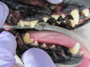
Pic 1: Left side of Lily’s teeth, just after her deep periodontal cleaning. X-rays showed both canine teeth had tooth resorption.
Tooth resorption is a painful process where the crown of the tooth is demineralized and ultimately breaks off. This can start in the crown (above the gum line) or in the roots of the tooth. If it is affecting the tooth roots, this does not appear to be a painful process and may be monitored with x-rays every 6 -12 months.
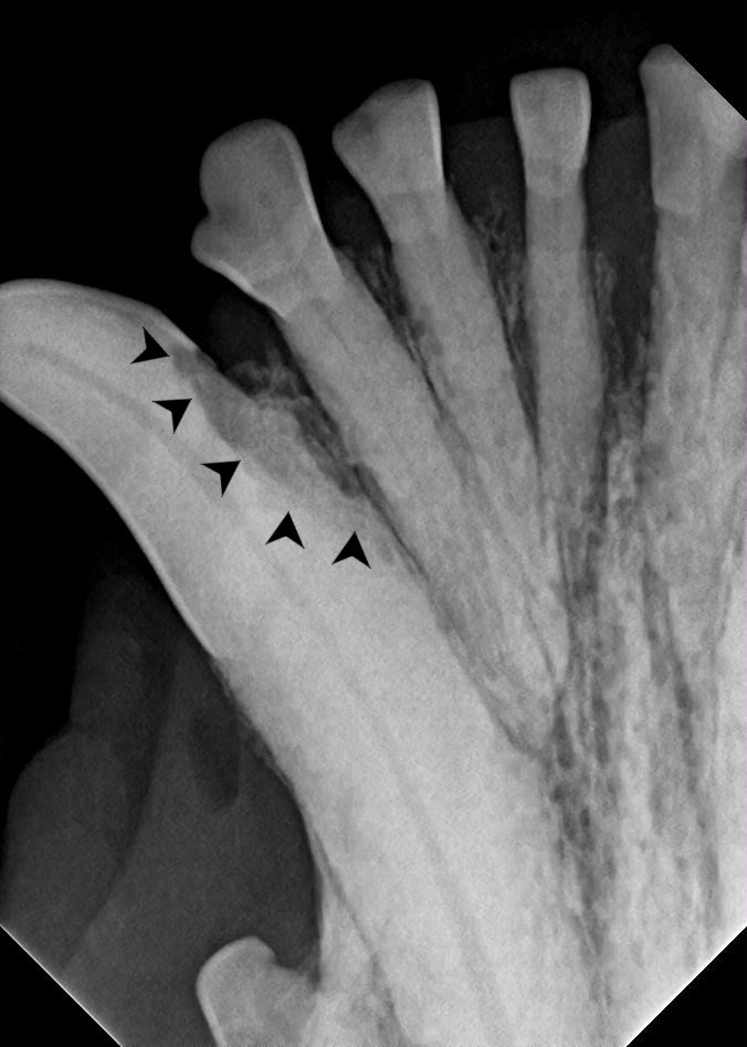
Pic 2: X-ray of lower left canine tooth. Black arrows highlight the area of the crown that is resorbing.
Pic 2: X-ray of lower left canine tooth. Black arrows highlight the area of the crown that is resorbing.
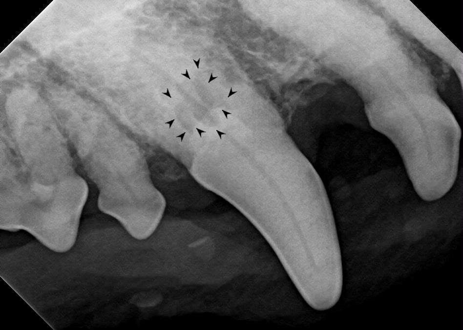
Pic 3: X-ray of lower left canine tooth that shows tooth resorption.
Pic 4: Upper left canine tooth. The black arrow shows the area of resorbing tooth root.
With our pets, the goal is for them to have a pain free mouth. Tooth resorption is a very painful disease. There is no treatment available besides removing the affected tooth to create a pain free mouth. Our dogs and cats do not need teeth to eat and eat better after having their painful teeth removed.
Lily is a great example of why routine dental exams and x-rays are so important. She was ultimately referred to a dental specialist and had 8 teeth extracted. Once her extraction sites healed, she had more energy and was back to her old self.
Oral pain in our pets is extremely hard to appreciate. Many times animals do not show signs that we recognize as pain. Animals will almost ALWAYS continue to eat even with extremely painful mouths. Animals with broken jaws will even eat! Many times they just “slow down” and it is attributed to old age when in fact it is chronic pain. Every animal needs a yearly dental prophylaxis under anesthesia with full mouth dental x-rays to ensure that we are not missing any of these painful conditions.

Additional Education
https://www.aaha.org/aaha-guidelines/dental-care/dental-care-home/
https://www.avdc.org/painanddisease.html
https://www.avma.org/public/PetCare/Pages/Pet-Dental-Care.aspx
http://www.toothvet.ca/PDFfiles/tr_dogs.pdf