Hip dysplasia (CHD) is a debilitating and painful condition that affects the hips of millions of dogs (and some cats!). Genetics plays a role, as do factors such as breed, hormones, growth rate, activity, body weight and age. Originally described in 1937, hip dysplasia has been studied over the years and while there are ongoing studies, to date there is no medical or surgical cure. As such, the veterinary focus has been directed towards early diagnosis, intervention and treatment of affected dogs, and encouraging selective breeding to decrease the risk of passing the genes along to offspring.
Dakota, a joyful female Golden Retriever, is our Pet of the Month for June. At three years of age, Dakota wasn’t showing any issues that her owner could discern and she was active and energetic. Dakota was potentially going to be bred and her owner wanted to be sure that Dakota’s hips were healthy. Dr. Robin Riedinger is one of only a few veterinarians in the greater Seattle area who is certified to perform PennHIP radiographs, the definitive diagnostic tool for evaluating whether a dog’s hips are at increased risk of developing hip dysplasia.
After a thorough physical exam, which was normal and didn’t detect any evidence of pain in the hip joints, Dakota was sedated and positioned for her radiographs. The images revealed significant laxity in both hip joints which is consistent with a diagnosis of hip dysplasia. This was disappointing news for Dakota’s owner, but now that she knows Dakota’s status, she can make appropriate decisions to help Dakota live the balance of her life as comfortably as possible. We appreciate Dakota’s owner being willing to share Dakota’s radiographs with the public and helping others to understand more about hip dysplasia.
The following images demonstrate the 3 views obtained during a PennHIP evaluation – the compression view, the distraction view and the hip-extended view. You can see in the distraction view just how far out of the socket Dakota’s hips are with mild distraction. This same change will happen when Dakota is awake and active. Eventually the laxity leads to bone remodeling (arthritis) and diminished joint function. This is painful and debilitating over time.
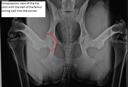
Compression View
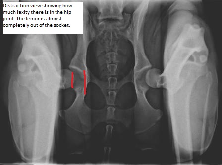
Distraction View
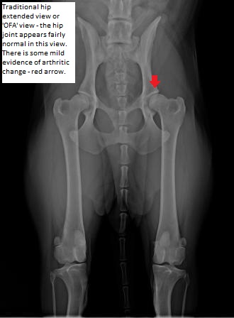
Hip Extended View
General guidelines for dogs at risk of developing hip arthritis include:
- Spay or neuter them as we don’t want to pass on the genetic risk to offspring
- Keep them lean and fit; regular exercise is great but limit activities that put undue stress on the hip joints (i.e. don’t have them catch frisbees in the air and land full force on their rear legs)
- Start them on a joint supportive food (such as Hill’s j/d or metabolic + mobility, Purina JM, or Royal Canin Joint Care)
- Don’t ignore any signs of limping or signs of pain/discomfort. Dogs don’t always complain in the same ways people do, so if they are, presume the pain is real and schedule a visit with your veterinarian
- Consider physical therapy options to build up the supportive muscles of the hip and rear legs, and learn about the various methods to help ease the symptoms associated with arthritis as it develops. Veterinarians certified in rehab medicine and acupuncture can also be helpful
- Discuss any surgical options with your veterinarian or a veterinary orthopedic specialist depending on the age of your dog and the degree of clinical symptoms
- Don’t withhold medications if your veterinarian has recommended them; your dog with thank you
WHAT IS THE PENNHIP METHOD?
Radiographs in general are an excellent method to evaluate the bones and joints, and can accurately tell which joints have developed arthritic changes (osteoarthritis). However, basic radiographs, and OFA (Orthopedic Foundation of America) images of the hips have failed to accurately diagnose and predict which dogs are at greater risk for hip dysplasia. In 1983 Dr. Gail Smith, a veterinarian at the University of Pennsylvania Veterinary School began researching better methods to determine which dogs were most susceptible, and in 1993 he established PennHIP.
Certified veterinarians follow strict protocols and all images are submitted and evaluated. PennHIP has been tested and shown to be scientifically consistent and predictive for the development of osteoarthritis. The method involves taking 3 separate views of the patient’s hips while the joint is positioned in an anatomically accurate position. Radiographs are taken under heavy sedation or anesthesia to ensure proper positioning and to eliminate any pain to those dogs who already have hip changes. Studies have shown that images can be taken in dogs as young as 16 weeks of age, and there is consistent correlation with their hip laxity at 2 years and 3 years of age.
The resulting images allow calculations to determine the laxity of the hip joint. Each breed of dog is compared with other dogs of the same breed; the individual patient is given a score to indicate where they fall along the spectrum. The database of over 100,000 dogs has allowed for a much more comprehensive assessment of the degree to which different breeds are affected. This helps pet owners know the risk for their individual dog, and it also allows breeders to make decisions to select for dogs with tighter hip joints, and thereby decrease the likelihood that offspring will inherit the trait. The ultimate goal – reducing the incidence of hip dysplasia and the subsequent development of osteoarthritis in dogs. That benefits everyone.
If you wish to have your dog’s hips evaluated, please call 206-528-1980 to schedule a consultation.
To learn more about PennHIP:
PennHIP website: https://antechimagingservices.com/antechweb/what-is-pennhip
PennHIP Brochure: http://ambientedepruebas.net/antechweb/pdf/AIS_PennHIP_Brochure_2015.pdf
Surgical Options for Dogs with Hip Dysplasia:
You need to discuss your own dog’s issues with a skilled veterinary surgeon to determine which, if any of these, are options for your dog.
- Juvenile pubic symphysiodesis – this must be done in young puppies; generally < 4 months of age
- Triple Pelvic Osteotomy (TPO) – the pelvis is realigned to allow the head of the femur to sit in the hip socket more normally; generally in dogs < year of age
- Femoral Head Ostectomy (FHO) – this procedure is typically done in dogs whose range of motion and comfort is significantly compromised. The head of the femur is removed which eliminates the ‘bone on bone’ pain of a diseased hip joint.
- Total Hip Replacement (THR) – replacing the diseased hip with an artificial joint, just as in people.
Local Veterinary Rehab Specialists:
- Sound Veterinary Rehab (Shoreline, WA) https://soundvetrehab.com/for-pet-owners/
- Seattle Veterinary Specialists in Kirkland, WA https://bluepearlvet.com/rehabilitation/