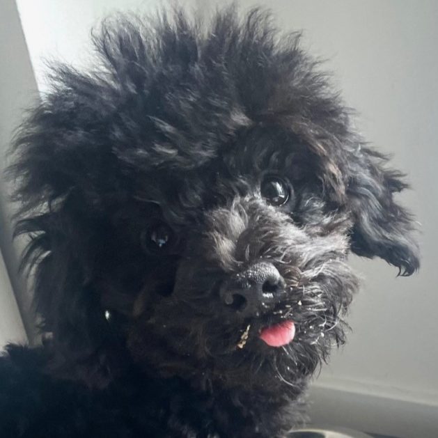
Toby is a 9.7# miniature poodle puppy with a sweet disposition. As is the case with most puppies, he has been learning new skills and recently learned how to jump up onto the furniture. He was enjoying himself one day several weeks ago, when his owner heard a loud thud. He had been trying to hop up on a chair and apparently misjudged. Initially he seemed okay, but started limping at home.
It can be hard to know if an injury is minor or more serious, especially because dogs and cats don’t always complain the same way people do. Toby was continuing to favor one hind leg on and off at home, so his owner scheduled an appointment with Dr. Riedinger at Hawthorne Hills Veterinary Hospital.
During the exam Toby was friendly and did not react to any portion of his examination. We palpated the hip joints, the knees, tarsus (ankle joint) and his patella (knee cap) and did not detect any fractured bones or specific painful areas. The only findings were the knee joint felt a little thicker on the right leg versus the opposite leg, and Toby wasn’t willing to put full weight on his hind leg when placed on the ground.
The best way to evaluate lameness that doesn’t resolve in a day or two, is to sedate the patient and get radiographs. Sedation is important so that we can get full relaxation and to position the area properly without causing pain to the patient. People can often hold a weird position for several minutes to get a good image, but pets just don’t understand. Sedation for imaging is also much safer for our team.
Growing puppies have multiple growth plates where two or more sections of bone are connected by cartilage. These areas are what allows the bones to grow longer or thicker over time, and as the pet reaches maturity, the growth plates close and are ultimately converted to bone. During the growth phase, these growth plates are vulnerable to injury and in some cases, malformations.
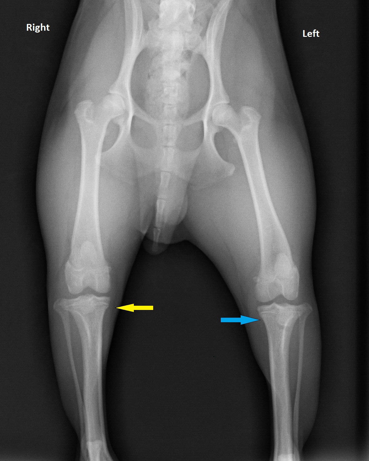
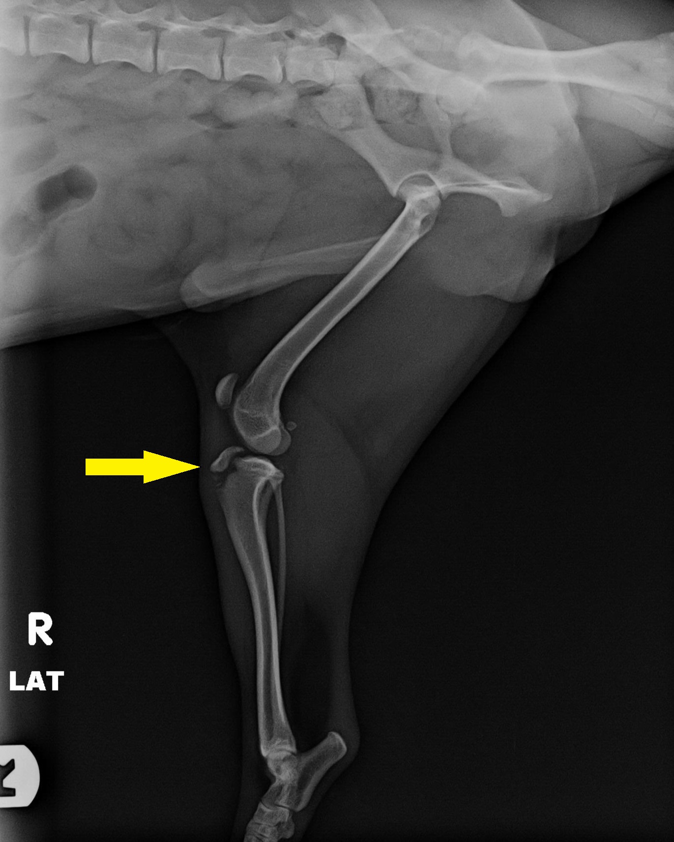
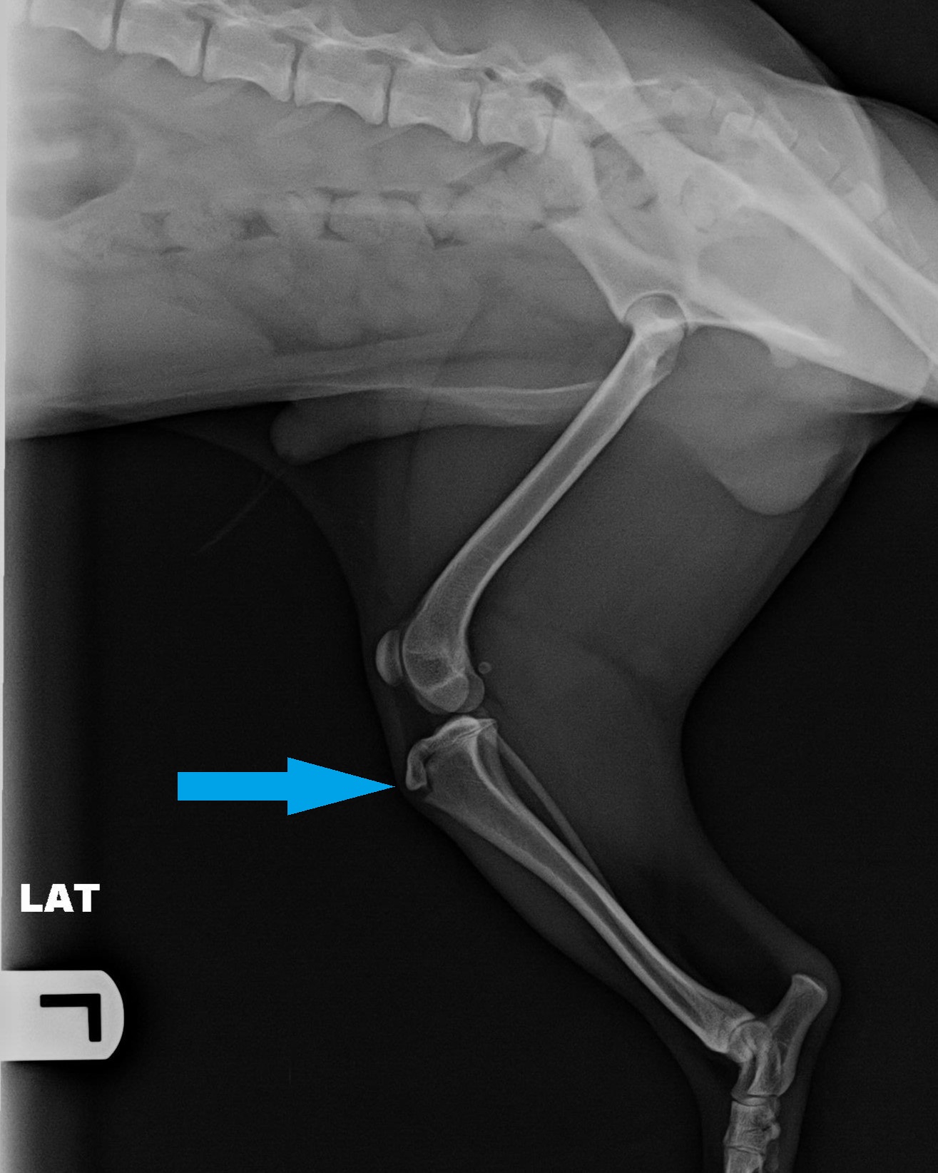
We sedated Toby and obtained radiographs of his hips and knees. The imaging revealed an avulsion fracture of the right tibial tuberosity. This is where the patellar tendon attaches to the lower leg bone. This type of injury is quite common in young dogs 5-9 months age, and frequently is related with some sort of jumping or falling injury and landing wrong. The strength of the quadriceps (thigh) muscle pulls the bone attachment away from the tibia.
The radiograph on the left was taken with Toby laying on his back. The yellow arrow points to the right tibial tuberosity (just below the knee joint) and the blue arrow points to the left joint. In this view the knee joints looks normal. The middle and right images show side views of Toby’s knee joint. You can see the patella (knee cap) just in front of and above the knee joint. The yellow arrow points to the avulsion fracture (displaced piece of bone) on the right hind leg; the blue arrow points to the normal appearance of the left tibial tuberosity.
Toby was referred to a local orthopedic surgeon for advice as to whether surgery was essential or could this potentially heal with conservative rest at home. Despite the displacement of the bone fragment, Toby’s patella was in its normal position and the knee joint was stable. Following discussions about the benefits of surgery and the risks of not pursuing surgery, along with costs, Toby’s owner opted against surgical intervention. Ultimately, Toby’s small size and weight, and the fact that he was already healing, made him a good candidate for conservative care. For a larger, heavier, more active breed of dog, surgically pinning the avulsed potion of bone back in place is generally required to avoid long term consequences.
Toby has been on a plan of restricted activity with no jumping for several weeks, then a gradual increase in daily walks over several weeks before returning to normal activity. His owner is monitoring Toby for any setbacks that suggest the injury is not healing well. We’ll continue to check in periodically and look forward to seeing Toby fully recovered.
