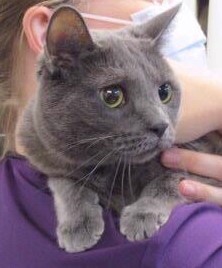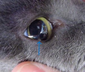
Claire’s family noticed that she had developed a small growth along the right lower eyelid and brought her in for an evaluation.
During the physical examination, Dr. Ravneet Kaur heard a gallop rhythm when listening to Claire’s heart. There was no murmur detected and while Claire had some mild tartar on her teeth, the rest of her exam was normal. This arrhythmia was concerning for a condition called Cardiomyopathy.
We are appreciative of Claire’s owner being willing to share her story for our Pet of the Month blog so that other pet owners can be educated. The news about a heart change was unexpected, and the road ahead may be challenging.
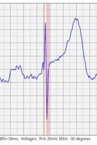
To gather more information, blood and urine samples were collected and an ECG was performed. The captured signal shows the electrical pulse wave as it moves through each of the heart chambers.
The ECG gives information about the heart rate, rhythm, strength, and timing of the heartbeats. Alterations in the ECG also give us information about the size of the heart and if there is underlying change. The ECG was sent remotely to a veterinary cardiologist who confirmed our findings, and diagnosed a ventricular arrhythmia. Claire was discharged while we were waiting for the lab results.
Concerned for her beloved pet, Claire’s owner brought her in the following day for a recheck. During the evaluation, it was clear that Claire was now breathing with more effort suggesting that her heart was not pumping blood effectively. In listening to her heart, the erratic arrhythmia was still present. Other diseases that might have been contributing to the heart changes were ruled-out.
Claire was referred to a local cardiologist for an echocardiogram. This is an ultrasound of the heart and allows the cardiologist to see the size, shape, and function of the different heart chambers. A cat’s heart is just like a person’s – there are four chambers that work in concert to receive unoxygenated blood from the body, circulate it through the lungs to pick up oxygen, and then send the newly oxygenated blood back to the body’s organs and tissues.
Depending on the type of heart disease, the problem can be with the valves (valvular heart disease is common in dogs and people), the vessels carrying the blood (coronoid artery disease in people), or it may be associated with the heart muscle itself. The normal sequence of the heart function happens in two stages – systole where the heart contracts and pumps blood out of the chambers, and diastole, when the heart muscle relaxes, allowing the chambers to fill with blood.
The echocardiogram led to a diagnosis of: ‘Unclassified Cardiomyopathy’ meaning the heart muscle is not contracting as it should but the left ventricle is not enlarged, therefore she doesn’t have Dilated Cardiomyopathy as it is typically defined. The cardiologist listed the changes present:
- Decompensated congestive heart failure
- Both left and right atria are severely enlarged
- Smoke present – at risk for thrombo-emboli
- Diminished systolic function
Per the cardiologist, ‘some boutique/grain free/exotic diets have been linked to nutritional mediated cardiomyopathy. The exact nature of this relationship is still being explored. Until we have answers, the best recommendation is to feed a food from a company meeting the nutritional guidelines the WSAVA (World Small Animal Veterinary Association) guidelines, and to avoid pea products (lentils, peas etc.) in the top 10 ingredients.‘
In Claire’s case the dilation of the atrial chambers means blood doesn’t flow properly through the heart with each contraction causing symptoms such as shortness of breath and decreased energy. Because blood can also pool in the heart chambers, it can also put Claire at risk for blood clots and sudden death. Signs of thrombo-embolism could include back leg paralysis, sudden onset pain, pale gums, cold rear toes/foot pads, and in some situations, sudden death.
Treatment plan:
- Furosemide to reduce fluid retention in the lungs and tissues by increasing urine production
- Pimobendan to help the heart contract more effectively, and dilate blood vessels to ease blood circulation
- Clopidogrel and Xarelto inhibit platelet clumping which reduces the likelihood of blood clots
- Taurine which is an essential amino acid for cats and a deficiency can contribute to cardiomyopathy in cats
- Switch to a prescription food designed to support heart function that does not have legumes in the formulation
Despite the challenges this diagnosis presented, Claire’s owner is committed to doing whatever is necessary to manage the heart disease and monitor Clair’s symptoms. She has worked out a system of giving medication and rewarding Claire for cooperation, and has introduced the special diet to support Clair’s heart health. Claire is back home enjoying her days, albeit with a bit more caution and rest. We are hopeful that the therapies will improve Claire’s heart function and keeping her purring for years to come.
Claire will be scheduled for a recheck echocardiogram and at that time, ‘if there is no improvement in her heart function, then diet related heart disease can be ruled out, and primary heart disease is more likely.’
If you have concerns about your cat’s heart function or suspect they may be experiencing symptoms of respiratory changes, please schedule an appointment with one of our doctors. We are here to help.
For more information:
- Pimobendan: Pimobendan for Cats: Overview, Dosage & Side Effects – Cats.com
- Dilated Cardiomyopathy in Dogs & Cats Dilated Cardiomyopathy in Dogs and Cats – Veterinary Partner – VIN
- Hypertrophic Cardiomyopathy Hypertrophic Cardiomyopathy in Cats – Veterinary Partner – VIN
- Cardiomyopathy in Cats Cardiomyopathy in Cats – Veterinary Partner – VIN
- Congestive Heart Failure in Dogs & Cats Congestive Heart Failure in Dogs and Cats – Veterinary Partner – VIN
- Diets and Heart Disease in Dogs & Cats Diets and Heart Disease in Dogs and Cats – Veterinary Partner – VIN
- WSAVA About (wsava.org)


