Dental care in dogs and cats is an important aspect of keeping them healthy. Home care is beneficial, whether toothbrushing, using VOHC approved dental wipes, adding dental hygiene products to their drinking water, or feeding a tartar reducing food. However, home care can’t replace the benefits of a professional assessment of your pet’s teeth under anesthesia.
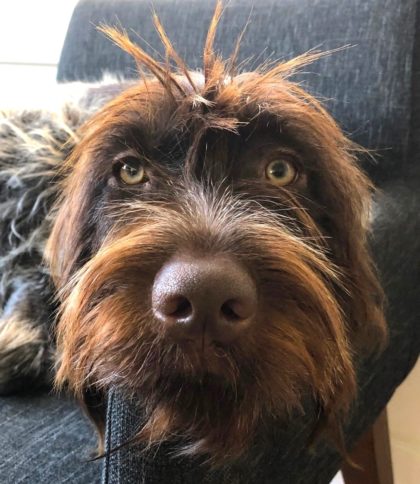
February is Dental Month and we would like to introduce you to Murray. She’s a lovely 5-year-old Wire-Haired Pointing Griffon.
Murray’s owners had noticed several areas where brown tartar was accumulating on her teeth which was confirmed during a physical exam. Despite the tartar, Murray only had mild gingivitis – that’s the redness along the gum lines. Some pets can have tartar buildup without gingivitis, and others can have gingivitis without much tartar. While we want to avoid significant tartar buildup, the gingivitis is the real issue. Inflammation along the gum lines can progress to infection below the gums and changes affecting the tooth roots. We obviously want to try and keep all teeth healthy for a pet’s lifetime.
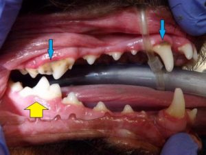 This picture shows some tartar buildup on the upper canine (fang) tooth and also on the largest chewing tooth in the back ( blue arrows).
While the other teeth have minimal tartar, the gums are mildly thicker in some areas – especially along the upper teeth.
Compare the upper gum line to the gum adjacent to the yellow arrow. The lower gum tissue is very thin, pale pink and healthy.
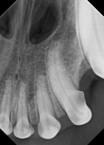
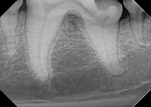
Here are several radiographs of normal teeth. The one shows the upper incisors – while the crowns of the teeth are smaller, the roots are quite long. The second image shows how large the roots are for the large cheek chewing teeth; in this case the lower left.
Earlier this month, Murray was admitted to Hawthorne Hills Veterinary Hospital for her first professional dental procedure.
Under anesthesia we took pictures, charted the teeth, and assessed them for crowding, pocketing and/or tooth damage. Radiographs of the teeth are particularly important as the crown is only 1/3 of the tooth – pet’s have long tooth roots that help anchor the teeth in place as that is what gives them strength.
Some of the things we evaluate on a radiograph are:
-
-
- Missing or unerupted teeth – teeth that could be buried below the gum line must be extracted to avoid the risk for dentigerous cysts
- Extra roots – not always a problem, but important to know if a tooth needs to be extracted
- Abnormal roots – some roots have bends or crooks which can pose a problem in the future, and make extractions very difficult
- Infection along the roots – loss of attachment to the underlying bone; some types of infection can be treated, others are too advanced and the tooth must be extracted
- Resorption or destruction of a tooth root – the body can resorb teeth for a variety of reasons which weakens the tooth and puts it at risk of breaking. When this happens, extractions are almost always necessary
- External resorption of the tooth root is not well understood. We don’t know the cause, but we do know that specific cells called osteoclasts invade the tooth root and eat away the normal structure. This is not the same as have infection, and extraction is the solution once there is insufficient root anchoring the tooth in place
- Fractured (broken) tooth roots – trauma to a tooth (ex: chewing on hard objects) can cause fractures; depending on the situation extraction may be needed. If only a portion of the crown is fractured, a root canal may be possible which saves the tooth
-
Murray’s radiographs revealed abnormality of 3 of her first premolars – those are the smaller teeth just behind the large fang (canine) teeth. The roots were being resorbed and when we probed around the tooth, all of these teeth were mobile – there wasn’t enough root to keep the tooth in place. There was no way to ‘save’ these teeth so they were extracted. We placed sutures to close the socket and while pets have to be on soft foods while the area heals, the sutures will dissolve over time. We are pleased to say that Murray took it all in stride and she is back feeling great.
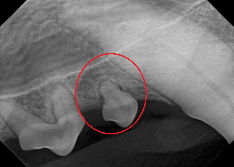
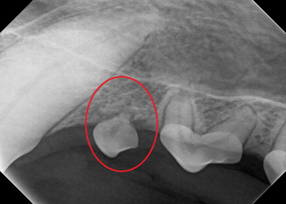
Here are photos and the corresponding radiograph to illustrate the root resorption happening with Murray’s teeth.
The first row shows the upper first premolars – the left is the right side; on the right is the upper left. The photos have an arrow pointing to the first premolar. The radiograph shows the tooth missing most of the root.


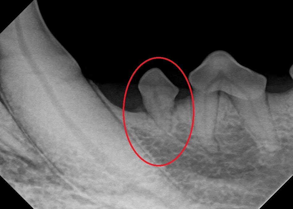
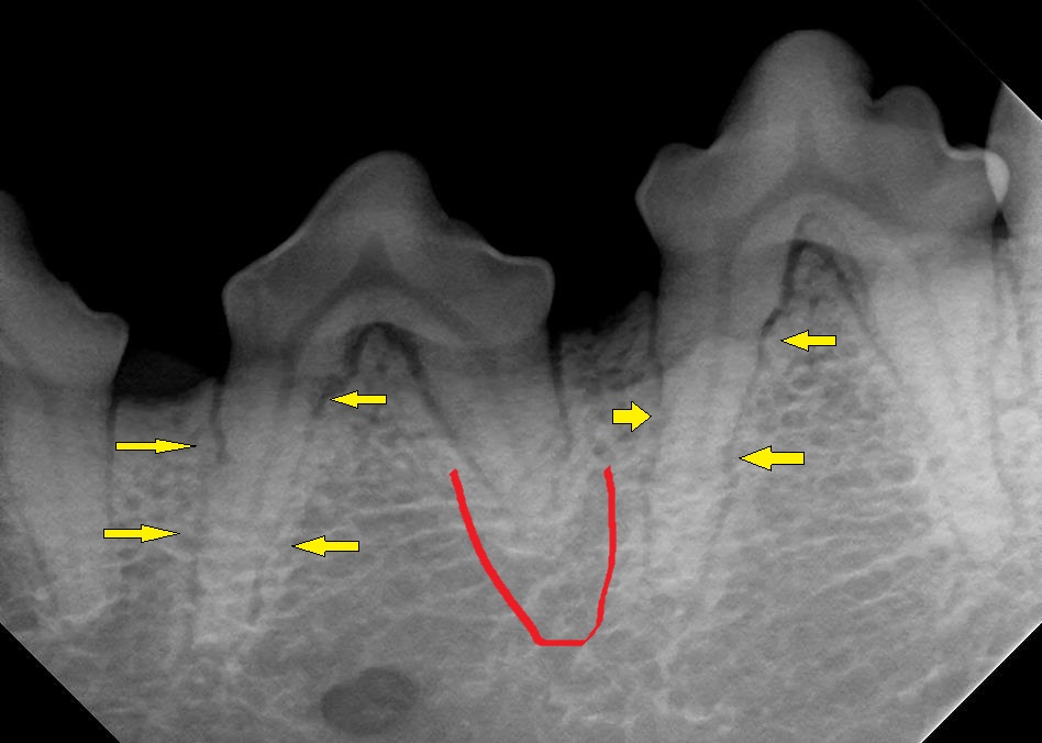
The second row of images, shows the left lower first premolar (left image) with the associated radiograph. The third image shows the normal right lower first premolar with the root delineated in red. The final image shows tooth resorption of the 3rd and 4th premolars on the lower left arcade. Yellow arrows point to areas of resorption; the root outline instead of being smooth, has a squiggly appearance. The red line shows how much of the distal root has already been resorbed.
Interestingly, Murray’s radiographs also showed early resorptive changes on a number of other teeth. We’ll monitor these teeth over the next several years; they may require extraction at some point.
If your pet has not had a professional oral exam or cleaning & assessment procedure under anesthesia, please call us for an appointment. Keeping your pet’s mouth healthy can add years to their lives.

