Our pet of the month for the month of September is Sylvie. She is almost 1.5 years old and her Owners adopted her in May from a local shelter. Sylvie is new to HHVH and saw Dr. Kaur for her first visit last month. Her owner’s mentioned she is doing well at home. During the exam we noticed a small bulge in her belly in the area of the belly button (umbilicus).The swelling appeared to be very soft and squishy below the skin. Sylvie was not bothered by palpating the swelling and the rest of her exam was normal. Based on the location we explained to Sylvie’s owner that it is an umbilical hernia and recommended surgical repair. Sylvie was scheduled for pre-operative blood work and surgery with Dr. Riedinger.
Hernias are the defects in the muscle wall where internal organs protrude through the abdominal wall. They can be caused by traumatic injury or congenital condition, meaning present at birth. In some cases they can develop after an abdominal surgery. Umbilical hernias are often seen in kittens because they occur as a result of umbilical rings not closing after the birth.
Umbilical hernias are usually painless and often a bulge is present in the belly button region especially when they are standing on all four legs. In some cases, it may not be apparent until maturity, when the cat has a condition causing increased abdominal pressure, such as obesity, trauma, or straining. Size of the hernia can vary and they may contain only fat or other internal organs. Most umbilical hernias do not cause any symptoms and are not an emergency.
Most of the time the hernia repair is done at the time of spay or neuter surgery so the patient has to go under anesthesia once. Sylvie was spayed by one of the shelters before she was adopted. However, she had a litter of kittens and developed severe mastitis (inflammation and infection of the breast tissues), so it is possible that the hernia was not noticed or the focus of surgery at the time was to get her in and out of surgery as quickly as possible. Prognosis is great after surgery.


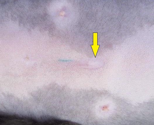
The first picture on the left, has a yellow arrow pointing to the umbilicus (belly button). The herniated fat has flattened and is not visible here. To the left of the yellow arrow you can see a green line; this tattoo is placed to indicate that the pet was spayed by the shelter. In the second photo, we’ve exposed the fat tissue at the site of the hernia. The green arrow points to the green tattoo. In the photo on the right, we’ve exposed the actual hole in the belly wall – red arrow. You can see how much fat tissue has pooched through the defect.
During the surgery it was noted the defect in the wall was small about 1 cm in the length and mostly contained abdominal fat. The abdominal wall was sutured after removing the herniated fat. In some cases if the defect is large, mesh is needed to close the defect and prevent internal organs pushing through the wall in future. Sylvie’s procedure went well and she recovered quickly post-surgery.
We saw Sylvie back for suture removal after 2 weeks. Her incision looked completely healed and we removed the skin sutures. Sylvie enjoyed her owner’s company during the recovery and here is a funny little story from that period from her owner.
“A few days after Sylvie’s surgery, I (Mom) was working from home. I only had 1 meeting all day and Sylvie decided that she wasn’t getting enough attention. She decided to sit on my closed laptop, and stared directly into my webcam not once, but TWICE during this 30 minute meeting, taking up my entire camera with her face. Thank goodness my coworkers got a kick out of it! “
Useful links for more information
VeterinaryPartner.com – Umbilical hernias in Puppies & Kittens
