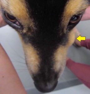
Our pet of the month for the month of July 2023 is Bailey. She is an 8-year-old active Australian shepherd. We received a phone call from Bailey’s owner that they were concerned about swelling on the left side of Bailey’s face that they had just noticed. They were not sure if the swelling just appeared but when they looked back at the pictures it is possible that it might have been there for a couple of weeks and increased drastically all of a sudden.
Dr. Ravneet Kaur examined Bailey and found there was prominent swelling on the left side of the face, under the eye face. No puncture wound, discharge, bleeding, or inflammation was noted on the skin around the swelling. When we examined Bailey closely, the swelling appeared to be under the gums at the level of the root of the upper 4th premolar tooth. Bailey was not painful, was eating and drinking without any difficulty. The rest of her physical exam was within normal limits.

 A fine needle aspirate was collected through the skin to get some answers as to the cause of swelling although it was discussed with her owners that most likely this was a dental/tooth problem.
Based on presentation and the location of the swelling, a tooth root abscess was highly suspected. Bailey would need an oral exam and dental radiographs under anesthesia. The other potential causes of facial swelling could be an injury, tissue abscess, an insect bite, or neoplasia.
The cytology report the next day revealed the presence of inflammatory cells. The report was not definitive but also suggestive of an abscess of the root of the biggest upper premolar. Bailey was scheduled for a dental procedure to look more thoroughly and obtain dental radiographs.
The most common tooth to develop a tooth root abscess is the large, upper carnassial tooth i.e., the 4th premolar. It was the same tooth Bailey was suspected to have an abscess. This tooth is prone to developing a slab fracture, in which a piece of the crown breaks away from the main tooth to expose the inner dentin and sometimes the root canal and giving access to the bacteria. Sometimes periodontal or severe gum disease causes the development of an abscessed tooth.
Dogs with tooth root abscesses may be asymptomatic in the early course of the disease. Some patients show subtle signs of pain like reluctance to chew, loss of appetite, and pawing at the face. Later in the course facial swelling, fever, lethargy, weight loss or other signs may develop if the infection spreads to other areas like the eye orbit.
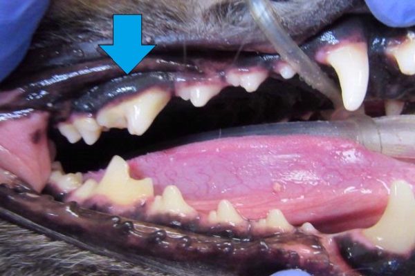
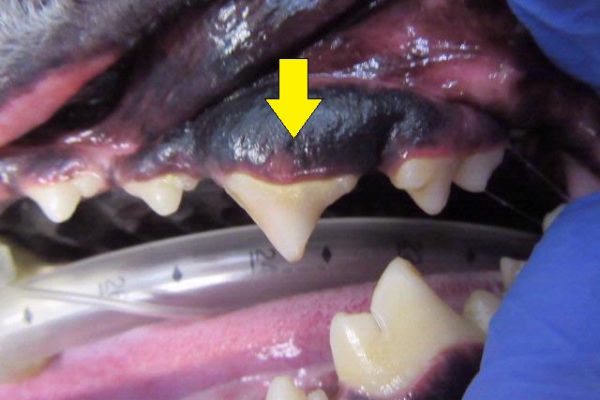
The photos below show the appearance of Bailey’s upper premolar 4 on the right (blue arrow) and on the left (yellow arrow). You will notice that the right side tooth has a typical shaped tooth with two crowns (the front crown tip is worn), whereas the left side tooth appears triangular, which is not normal. It wasn’t until Bailey was under anesthesia that we were able to get a close up look at the full tooth.
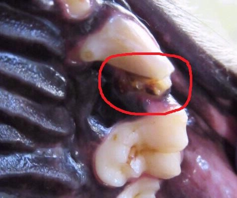
Under full anesthesia, we can get a close up look at the teeth and gums. The oral exam may reveal swelling or inflammation of the gums, fractured, or discolored or loose teeth, and purulent discharge in the oral cavity or face if a fistula is formed. In Bailey’s case, when we examined the tooth under anesthesia it was noted that the crown ( part of the tooth that is exposed and visible above the gum line) was missing a portion and appeared abnormal in shape.
Dental radiographs are very important to confirm the diagnosis. Typically, the radiolucent area (halo) surrounding the apex of the tooth root is visible on intra-oral dental radiographs. Some other changes are the thickening of the periodontal ligament, swelling around the affected root, and abnormalities of the pulp cavity. In some cases, infection of the bone is also evident.
Complicated cases may need advanced imaging techniques like CT scans to clarify what is happening, especially if there is not an obvious tooth root abscess. Bone disease, other types of soft tissue swelling, nasal disease associated with it, the extent of bone loss, extension of the infection to eye orbit or neoplasia.
Most tooth root abscesses are treated with extraction of the involved tooth, particularly if the crown of the tooth is damaged and cannot be repaired. In some cases, endodontic procedures like root canal, done by a dental specialist may be used depending upon the extent of damage and severity of the infection. Antibiotics can be started while waiting for the dental procedure but are not sufficient to treat this condition.
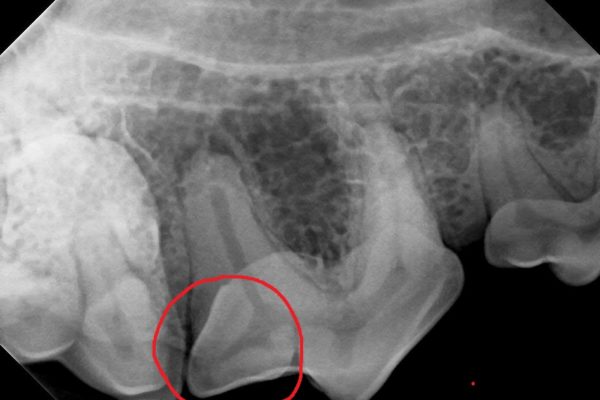
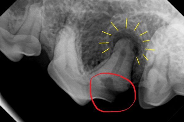
Once Bailey was under anesthesia, we were able to see that her tooth was fractured and not just malformed. The photograph shows the missing portion of Bailey’s tooth – circled in red. The radiograph on the left shows the normal upper right premolar 4 for comparison with the left upper premolar 4. The red circle outlines the same location on each tooth.
The radiograph confirmed the presence of an abscess at the caudal root. The yellow lines outline the area of bone lucency which is consistent with an abscess at the caudal root. Because a portion of the crown was missing, this tooth could not be treated with a root canal procedure. After discussing the treatment options with the owner, Bailey’s abscessed tooth was extracted.
Post-extraction intraoral radiographs were taken to ensure all of the roots (there are 3 large roots on this tooth) are removed. Any of the root fragments remaining could develop an abscess again. The gum was sutured closed and Bailey was discharged with medications to keep her comfortable while she was healing.
Bailey did well after the procedure and was sent home with meds and instructed to have a soft diet for several weeks. Fourteen days later, at her oral health recheck, the facial swelling was completely gone and the site of extraction appeared to be healing well. Once the gums are completely healed dogs can resume their normal diet and activity.
It is a very common concern among pet parents how their dog is going to chew after the extraction. In fact, most dogs eat well once the painful tooth is removed. Dental care is an integral part of your pet’s overall health. Here are a few things we can do to prevent tooth root abscesses in dogs:
- Schedule routine health check-ups
- Follow up with regular professional dental evaluation and cleaning under anesthesia
- Regular tooth brushing and oral care at home can help to prevent periodontal disease
- Minimize the opportunities for dogs to chew on hard objects to avoid tooth fractures – avoid cow hooves, antlers, hard inflexible toys, items like rocks
Useful links for more information.
-Dental care for pets- https://veterinarypartner.vin.com/default.aspx?pid=19239&id=4951294–
-Safe toys for puppies-https://veterinarypartner.vin.com/default.aspx?pid=19239&id=10282231

