
Jinddu recently had her teeth cleaned and has volunteered to share her procedure with you. Dental care is an important part of keeping healthy and a professional cleaning under anesthesia is needed every year or two. The frequency depends on several factors – home care, conformation of the mouth and teeth, genetics, and any injuries that may affect the teeth or gums.
Bacteria and plaque build up within hours of eating; that’s the fuzzy surface debris that you can feel with your tongue. As is true in people, brushing your pet’s teeth is one of the most effective tools to remove this film. In animals, the plaque becomes hardened into tartar after 24 hours and can only be removed by scraping it off the tooth surface. This accumulation of bacteria and minerals and pushes against the gum line as the material builds up. The tartar can have various colors from light tan or yellow to dark brown, black or green and frequently has a very strong unpleasant odor.
Jinddu, a Korean Jindo, moved to Seattle with her family from South Korea. She just turned 2 years old and had a mild amount of tartar already accumulating on her teeth. In addition, she appeared to be missing several teeth. In younger dogs, if teeth are not visible the question is always, did the tooth not erupt above the gum line correctly, or did the tooth just not develop? Dental radiographs as essential to know if there is a problem or not.
In advance of the procedure, we drew blood from Jinddu to ensure she was okay to undergo anesthesia. We also sent home some Gabapentin to be given at home the night before and the morning Jinddu was admitted to the hospital. This helped her to be more relaxed and comfortable during her stay. After arrival, Jinddu was weighed, had her vitals (temp, heart rate and respiration) checked and then was examined by Dr. Riedinger. All of this is to make sure that nothing had changed from Jinddu’s last visit.
There is no effective or painless way of fully evaluating all of a dog’s teeth above and below the gum line without anesthesia. A full COHAT – comprehensive oral health assessment and treatment, encompasses a number of steps. Teeth have multiple visible surfaces: the front (mesial), back (distal), the side toward the lips (buccal), the side towards the tongue (lingual) in addition to the chewing or occlusal surface. Depending on the tooth, there are typically one to three roots that anchor the tooth in the bone, although variations can occur. During a dental procedure all tooth surfaces and the oral cavity are evaluated. We:
- Take photos of the before and after
- Chart – record the amount of tartar, the level of gum inflammation, and the depth of any pocketing
- Document any changes associated with the tooth – chips, wear, fractures, crowding
- Identify any missing teeth or extra teeth
- Evaluate all of the oral tissues – under the tongue, the roof of the mouth, the tonsils, the pharynx and the epiglottis for any signs of abnormal change
- Get radiographs of all arcades – left and right, top (maxilla) and bottom (mandible) and record the findings
Jinddu had a numbing cream applied to her leg before we placed an intravenous (IV) catheter. Then we used a combination of drugs to get her asleep. She was intubated for the delivery of oxygen and anesthetic gas to keep her asleep during the procedure. During every anesthesia we have a dedicated skilled assistant who helps the technician and veterinarian record our findings. In addition, we utilize monitoring equipment which tracks heart rate, ECG, blood pressure, oxygen saturation, exhaled CO2, and temperature to aid us in maintaining an ideal depth of anesthesia. Every patient is also placed on IV fluids to help maintain heart and blood pressure and to ensure they are adequately hydrated. Some patients require pain medications and/or local anesthetic blocks to numb areas in the mouth that might be painful. That is always necessary if extractions are needed.
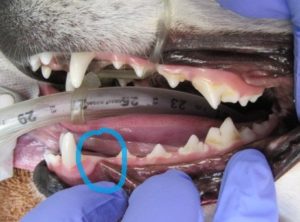
Blue circle shows the missing 1st lower left premolar
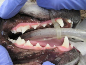
Right side teeth – all are present
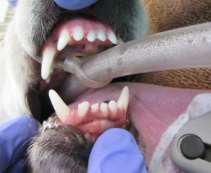
Lower incisors – one tooth is missing
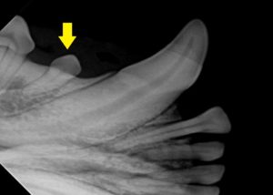
Yellow arrow shows the Right Lower 1st Premolar
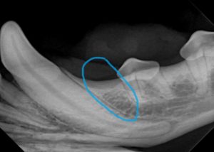
Blue circle shows where the Left 1st premolar is missing
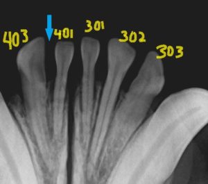
There are only 5 of the normal 6 lower incisors. #402 is missing and #403 has moved toward the center
The teeth are hand scaled to remove the large tartar accumulations. Then we use an ultrasonic scaler and finally polish all teeth. We did confirm that she was missing two teeth and two other teeth had extra roots. Neither of these changes required treatment.
While missing teeth are just a cosmetic issue, teeth are not meant to be trapped below the gum line. Buried teeth need to be extracted as they can increase the risk for developing a dentigerous cyst which can be quite serious if left untreated. Broken teeth, where the crown is gone but the roots remain pose another problem. In almost all cases the roots should be extracted.
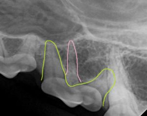
The green line shows the normal 2 roots. The pink the extra root.
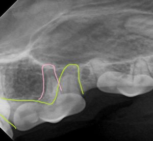
The green line shows the normal 2 roots. The pink the extra root.
We did uncover one interesting change. Two of Jinddu’s upper premolars had 3 roots versus the normal 2 – this is only a concern if that tooth needs to be extracted in the future because all roots need to be removed, and extra roots influences the extraction technique.
Jinddu’s teeth were quite healthy and only required cleaning and polishing. We applied OraVet Gel at the end which helps slow the build up of plaque on the tooth surface. She was allowed to wake up slowly and once recovered enough, she was given a small bit of food. She ate and then took a nap until her family came to pick her up.
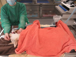
Here’s are pictures of Jinddu’s procedure:
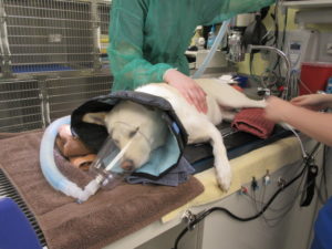
On oxygen and getting blood pressure and ECG connected
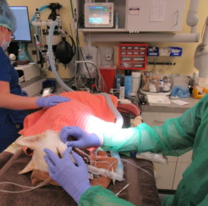
Fully under anesthesia – charting the teeth and recording the findings
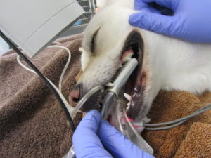
Placing the dental radiograph sensor
Taking a dental radiograph
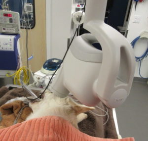
Procedure is done, being monitored until wakes up and can be extubated
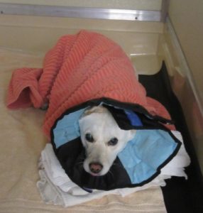
All snuggled in and waking up calmly
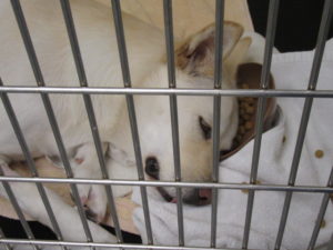
Taking a nap after eating a snack – head is in the food bowl
Links for additional information:
- https://hhvh.net/videos/brushing-your-dogs-teeth/
- https://hhvh.net/videos/brushing-your-cats-teeth/
- HealthyMouth.com – home care products that work
- Why Your Pet Needs Anesthesia for Oral Care