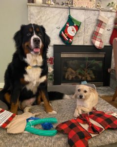
Clover is a one-year-old Bernese Mountain dog. Her owners decided to get her on a whim last year and she has been an important part of the family since. She’s relatively on the smaller side for a Bernese Mountain dog, but still packed with lots of fun. She’s very social and has been a welcome sight every time owners come home from work. She has a “big” sister, Daisy, who has been her best friend and also favorite playmate.
Discussing Clover’s case here we wanted to shed light on joint issues in young dogs. During Clover’s first veterinary visits she seemed to be growing appropriately and didn’t have any specific problems. Then, in July, Clover was seen at our hospital for eye discharge and soft stools. The owner mentioned about her gait being different. On exam she was very sensitive and painful to touch in the hips, and her back legs had an abnormal stance. We noted that her hock joints were hyperextended, meaning a straighter stance than angled which indicated problems up higher in the knee, hips or spine.
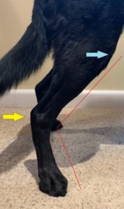
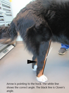
You can compare the photos below showing a normal rear leg stance and Clover’s stance. The yellow arrow points to the hock (ankle) joint, and the blue arrow to the location of the knee. The red line shows the normal angle between the knee, hock and paw in dogs. You can see how straight Clover’s rear leg stance was; she was shifting the weight off of her painful hips and changing the orientation of her lower leg bones.
 Clover was sent home on pain medication and referred to an orthopedic specialist. We had a high suspicion of a hip abnormality given her young age and breed. After the evaluation and radiographs were done at the specialty center, it was confirmed that Clover had bilateral hip dysplasia, meaning the changes involved both hips.
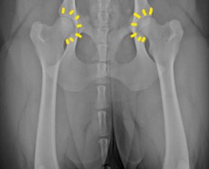
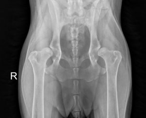
Hip dysplasia (HD) is abnormal growth and development of a dog’s hip joint. It occurs commonly in large breed dogs such as Labrador retrievers, German Shepherds, Rottweilers, and Saint Bernard, but it can occur in dogs of any breed and size, and even in cats. The cause of hip dysplasia is multifactorial, genetics being single biggest risk factor along with nutrition, and excessive, rapid growth. The hip joint is a ball and socket joint. The acetabulum, the socket portion of the hip joint, should be a deep ‘C’ shape. [Note the yellow lines outlining the normal hip socket].
The ‘ball’ portion is the head of the femur (upper rear leg bone), which should fit smoothly into the socket. If the socket is not shaped correctly, there is excessive hip joint laxity or looseness, along with a change in how the dog stands or walks. In turn, the laxity is responsible for symptoms of hip pain, lameness, discomfort, and progressive joint changes. The long-term response to this joint laxity is the progressive loss of cartilage, development of scar tissue around the joint, and the formation of osteophytes (bone spurs) around the ball and socket.
 The symptoms may be presented at a young age between 6-12 months due to hip laxity, or after maturity and into middle age, due to secondary arthritic changes in the joint. The main signs include lameness (mainly in back legs) which can be more pronounced after the exercise, reluctance to jump, running with a ‘bunny hopping’ gait, shifting weight to the forelimbs, and hip pain.
In Clover’s case, her initial puppy exams done at different veterinary facility were normal with no reported joint issues. Her joints palpated normally in May during her first exam at Hawthorne Hills Veterinary Hospital when she was 8 months old. By July, Clover was showing symptoms of pain and discomfort and a diagnosis was confirmed in August, after radiographs were obtained with the orthopedic specialist.
 The diagnosis is made by special palpation of the joints that determine abnormal joint looseness. Oftentimes this technique requires sedation or anesthesia in symptomatic dogs because it is painful to manipulate the joints. Radiographs are essential in diagnosis and planning the treatment.
Compare Clover’s radiograph of her hips to the normal hip image above. The acetabulum on both sides was very shallow and the head of the femur already out of the socket. Her diagnosis was clear based on the imaging.
However clinical signs do not always correlate with radiographic changes, and routine images may not accurately show or be predictive of hip dysplasia in very young ages. Dogs with symptoms, may benefit from specially positioned hip x-rays which focus on the demonstration and assessment of hip laxity. The PennHIP method is a way to assess puppies as early as 4 months of age, as well as dogs and cats of any age. This technique has the significant advantage of early diagnosis in breeds that are at risk and accurately predicts the risk for a puppy developing hip dysplasia. Early diagnosis widens the treatment options and prevents development of arthritis in joints. Special training and equipment are necessary to perform the PennHip radiographs. Dr. Riedinger is certified and has been performing this technique for many years.
Depending on your pet’s age, physical condition, and degree of hip pain/lameness, there are several surgical treatment options. Puppies diagnosed early, around 12-16 weeks of age, have the chance to be treated with a minimally invasive surgery called ‘Juvenile Pubic Symphysiodesis.’ Slightly older young dogs may be candidates for a ‘Triple Pelvic Osteotomy’ surgery where cuts are made in the pelvis to rotate the bones and reduce the laxity and movement in the hip joint. Unfortunately, once there is arthritic change in the hip joint, neither of these procedures can be performed.
Once arthritis is present, there are only two options to reduce the pain: 1) total hip replacement surgery or 2) a femoral head ostectomy, which is considered a salvage procedure to eliminate the bone-on-bone contact and pain.
The evaluation with the specialist determined that Clover would do best with a total hip replacement. She had her first surgery on the left hip, in October. She is recovering well and slowly resuming her activities. As her other hip is also affected, it is recommended to have the same treatment for the other hip, which will likely be done in a month or two.
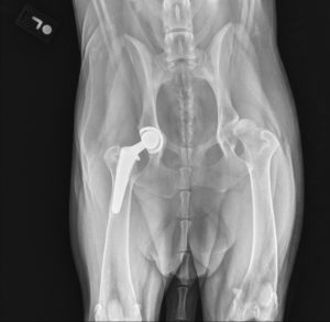
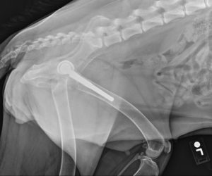 These images show the artificial hip joint implanted – a nice deep ball and socket and proper orientation of the leg and pelvis. This is the same procedure that is done in people.
We are glad to know that Clover is getting the care she needs. She will need rehab over the next several months as she adjusts to her new hips.
However, she should be pain free and able to enjoy a active life with her family into the future.
 If you want to know more, here on some links:
Links to PennHip information:
-
- PennHIP Brochure for Pet Owners
- PennHIP Fast Facts – Behind the Research fast facts
- PennHIP Video – the process video